
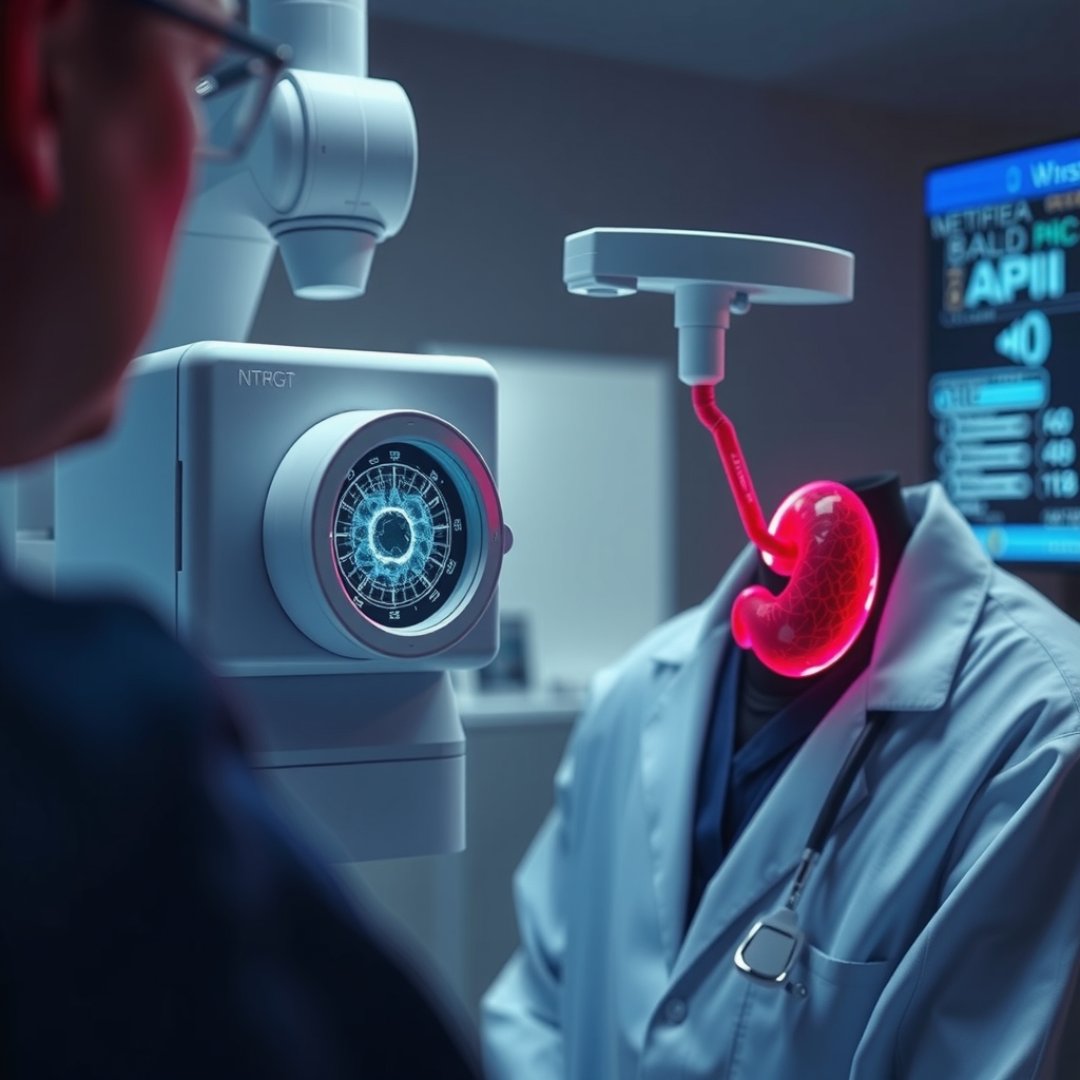
Pancreatic cancer remains one of the most challenging malignancies to treat, largely because it is often diagnosed at an advanced stage when therapeutic options are severely limited. Breakthroughs in biotechnology are now pivoting toward non-invasive methods, specifically focusing on pancreatic cancer early detection via electrical cell profiling. By measuring the unique dielectric properties and electrical signatures of circulating cells in the bloodstream, researchers can distinguish malignant cells from healthy ones with unprecedented sensitivity. This diagnostic paradigm shift allows for the identification of anomalies long before physical symptoms appear or traditional imaging reveals tumors. Understanding this technology is essential for patients and clinicians alike, as it represents a significant leap forward in improving long-term survival rates through timely, actionable clinical intervention.
The Science of Electrical Profiling
How Dielectric Spectroscopy Works
At the core of this innovation is the analysis of cellular electrical properties, often referred to as dielectric phenotyping. Every cell in the human body possesses a specific electrical signature determined by its membrane composition, internal organelles, and overall structural integrity. Malignant cells, particularly those shed by a tumor, exhibit distinct electrical abnormalities compared to their healthy counterparts.
Identifying Malignant Signatures
- Membrane capacitance: Cancerous cells often display altered charge distribution across their outer layers.
- Cytoplasmic conductivity: Metabolic changes in tumor cells shift their internal ionic environment, detectable via high-frequency electrical pulses.
- Morphological screening: Subtle physical changes in cell volume correlate with shifts in impedance measurements during flow-based analysis.
Advantages Over Traditional Diagnostics
Overcoming Imaging Limitations
Traditional diagnostic tools like CT scans or endoscopic ultrasounds often struggle to detect micro-tumors in the pancreas. Because the pancreas is located deep within the abdominal cavity, early-stage lesions are frequently obscured. Electrical cell profiling bypasses these anatomical hurdles by sampling blood, which acts as a liquid biopsy. This approach is minimally invasive, repeatable, and capable of capturing real-time molecular data that imaging simply cannot provide.
Reducing False Negatives
By focusing on the electrical fingerprint rather than visual shape, the technology reduces human error in interpretation. Artificial intelligence algorithms process the massive datasets produced by these sensors, identifying patterns that would be invisible to the naked eye, thereby significantly lowering false-negative rates in high-risk patients.
Clinical Impact and Future Potential
Improving Patient Survival
The primary benefit of adopting this technology is the ability to shift the timing of diagnosis. When a pancreatic malignancy is caught at stage I or II, surgical resection is much more likely to be successful. Increasing the number of patients eligible for curative surgery could fundamentally alter the mortality statistics currently associated with pancreatic cancer.
Scaling the Technology
Future applications include:
- Integration into routine annual blood screenings for high-risk populations.
- Monitoring post-treatment patients for early signs of disease recurrence.
- Developing portable chip-based sensors for point-of-care diagnostics in rural or underserved clinics.
Frequently Asked Questions
Is electrical cell profiling invasive?
No, it is a non-invasive procedure that only requires a standard blood draw to collect the circulating cells needed for the electrical analysis.
How accurate is this method compared to a biopsy?
While it does not replace a definitive biopsy, it serves as a highly sensitive screening tool that can identify patients who need urgent follow-up diagnostic procedures.
When will this technology be available in hospitals?
The technology is currently in advanced clinical research and pilot phases; broader hospital integration is expected as regulatory bodies finalize validation protocols.